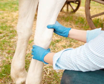
Thermography, or thermal imaging, is a simple, non-invasive way of detecting injury or potential injuries in horses.
What does it show?
When used correctly thermography can pinpoint muscle pain, stresses to the musculoskeletal system after extreme exertion or a fall, and nerve injury which may be implied by areas of atrophy (reduced circulation).
Preparation is key
Prior to being photographed, a horse must be unrugged, free of leg bandages, out of sunlight and away from drafts – all variables that can affect findings.
Thermographers will also take dedicated notes of the surroundings to aid them when deciphering the readings.
A full body scan involves 50 to 60 images of the horse, both before and after exercise, to build a complete picture of the horse’s physiology.
Pinpointing pain – how does it work?
Horses emit infrared radiation as part of a normal physiological process, and some of that heat passes into the air as heat photons.
A thermal imaging camera measures the heat photons emitted from a horse, and converts them into electrical impulses that are then displayed as coloured images on a monitor.
This map of the horse’s body temperature is called a thermogram and subtle abnormal temperature changes can be identified.
Why do temperature changes indicate injury?
Hot spots can indicate inflammation or increased circulation such as strains and bruising. They are generally seen in the skin overlying an injury.
A cold spot refers to a reduction in bloody supply usually due to swelling, scar tissue or nerve damage.
Data is interpreted through patterns. It is the identification of these thermal patterns determined by the horse’s anatomy, physiology, surface contours and the ambient temperature that is key to success.
Don’t miss the latest issue of Your Horse Magazine, jam-packed with training and veterinary advice, horse-care tips and the latest equestrian products available on shop shelves, on sale now.