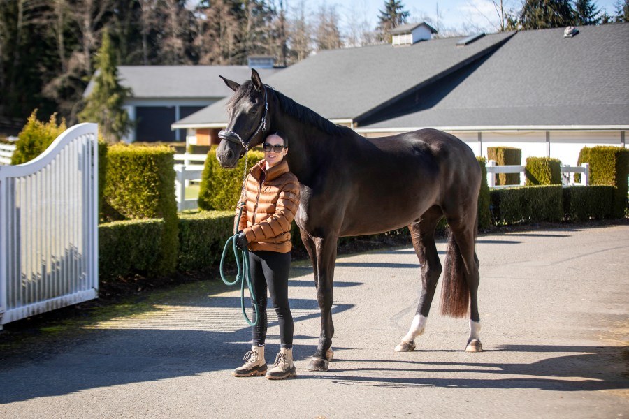
A four-year-old Oldenburg mare has successfully undergone highly specialised foraminotomy surgery – a minimally invasive procedure to relieve spinal nerve compression – in the USA.
Frida was one of the first patients to have the surgery at the University of California (UC), Davis, near Sacramento in December.
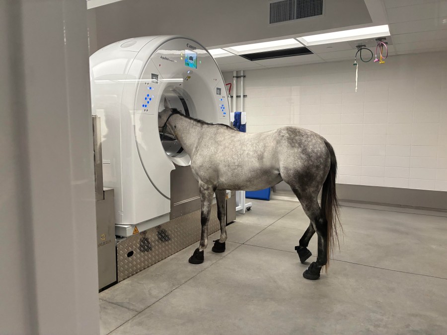
UC Davis has recently welcomed Dr Carter Judy as clinical professor of equine surgery as well as obtaining a new large bore equine CT scanner, allowing the UC Davis William R. Pritchard Veterinary Medical Teaching Hospital (VMTH) to perform the surgery.
Foraminotomy surgery is performed on horses with cervical foraminal stenosis – a narrowing of an intervertebral opening where the limb nerve root exits the spinal column.
The condition can be caused by arthritis, bone spurs, disc herniation, or thickened ligaments. It places compression on the nerve resulting in pain and numbness that can develop into lameness and behavioural issues such as headshaking.
New equipment
In order to diagnose the condition, which generally occurs near the base of the neck where the cervical and thoracic vertebrae meet, a large bore equine CT scanner is required, equipment which few equine hospitals have.
Previously, UC Davis only had a traditional CT scanner which meant it could not image beyond a horse’s head and distal limbs.
Osteoarthritis of multiple facet joints had caused foraminal stenosis in Frida, who is from Washington, which had resulted in hind-limb weakness, range-of-motion and coordination problems.
“Thanks to our recent acquisition of a large bore equine CT scanner, we’re able to achieve more accurate diagnoses of conditions such as foraminal stenosis,” said Dr Judy, at UC Davis.
“This condition causes a pinching of nerves as they come out of the spinal cord. In Frida, her foraminal stenosis resulted in behavioural and lameness abnormalities.
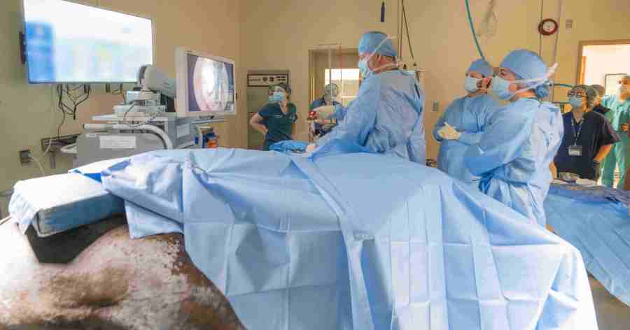
“She responded well to corrective surgery, which includes burring of a larger opening for the nerve, relieving the pressure on the nerve root. At Frida’s two-month recheck appointment, she showed a marked improvement in her clinical signs. I was very encouraged by how she looked.”
Joe’s story
The new scanner was also used to remove a sialolith (a hardened mineral deposit) from a 20-year-old American Quarter Horse.
Joe Juice, who is a member of UC Davis Center for Equine Health’s (CEH) teaching herd has a history if sialolithiasis having had a sialolith removed from the side of his face in 2021.
When a visible bump on his cheek appeared last summer, the team at UC Davis suspected he had another sialolith.
Joe Juice was one of the first models for the new equine CT scanner. His scan revealed a large, oval, mineral opaque structure measuring approximately 2cm x 3cm x 6cm within the soft tissues of his cheek, consistent with a sialolith.
‘Model patient’
“Joe Juice was a model patient for our standing CT training,” said Dr. Mathieu Spriet, director of imaging services at the VMTH.
“His sialolith was definitively diagnosed with the standing CT, giving surgeons a clear and concise assessment of the growth. This equipment’s ability to determine injuries and disease without full anaesthesia is already proving to be tremendously valuable for our patients.”
A few weeks later, Joe Juice had his sialolith removed and has since returned to CEH and resumed normal activity.
Images © UC Davis.
Related content
- Peritonitis in horses, which can look a lot like colic, explained by a vet
- Impaction and gassy colic in horses explained by a vet
- Inside a horse’s digestive system and how to keep it healthy
- Gastric ulcers in horses: why the time of day you ride and feed is so important
- 10 golden rules of feeding for a happy and healthy horse