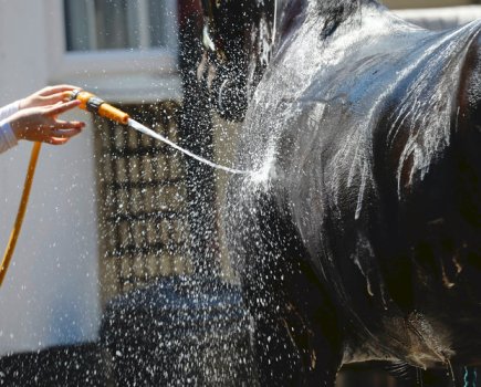
Thermal imaging works by building up a picture of your horse’s circulation and thermal pattern in any area of his body in a simple, non-invasive, stress-free way. It enables the identification of problem areas, known as ‘hotspots’ or ‘coldspots’.
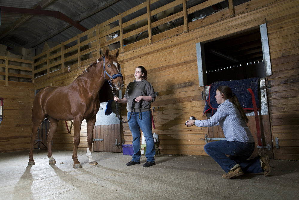
Photos are taken using the thermal imaging camera
How it works
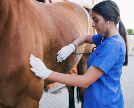
Thermal imaging looks at circulation and blood flow. The whole process involves an infrared camera which detects circulation changes in your horse’s body, with readings taken from a range of sites to allow for comparison. When trauma or injury occurs, a chain of chemical reactions take place which increase blood flow to the area. Circulation and blood flow dictate the thermal pattern and the infrared camera can then detect inflammation, which occurs as a ‘hotspot’. Abnormally cool areas can also be detected and indicate issues such as poor circulation, arthritis, nerve damage, oedema or atrophy (muscle wastage).
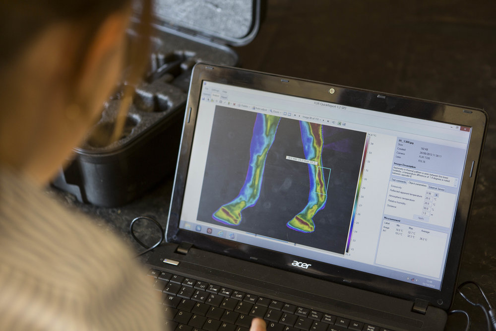
These can then be uploaded and assessed on a computer
What is it used for?
Thermography is proving to be an invaluable tool in the detection of issues such as:
– Ligament and tendon damage
– Back pain/kissing spines/ saddle fit issues
– Bone injuries/splints/stress fractures/arthritis
– Sacroiliac pain/injury
– Laminitis and navicular syndrome
– Hoof abscesses or imbalance
– Dental issues
– Muscle strain and atrophy
– Nerve damage
As well as the detection of injury and illness, thermal imaging can also be used as a fantastic and effective monitoring tool. When working to an intense training and competition schedule or bringing your horse back into work after time off, thermography can also help detect any niggles early. Ask your thermographer for a baseline full body scan to work out what’s healthy and normal for your horse, then as your workload increase, have your horse scanned at intervals to check out what’s going on and how he’s coping with the pressure. Monitoring a known injury site will also provide you with the information needed to see what lies beneath and to ensure that nothing worsens.
Research conducted in America has proved this by showing that thermal imaging can detect changes in structures – such as flexor tendons – up to three weeks before your horse shows any clinical signs of lameness. If injuries can be detected early enough, appropriate management changes can be put into place to save him from further damage – think of it as having the beauty of foresight!
Don’t miss the latest issue of Your Horse Magazine, jam-packed with training and veterinary advice, horse-care tips and the latest equestrian products available on shop shelves, on sale now.