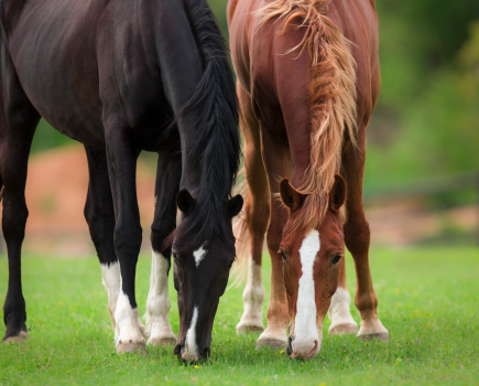
Legs that are continuously wet or muddy with or without mud fever are the perfect environment to incubate a case of cellulitis.
This is why a common complaint that I hear from owners worried about their horse’s health (especially in winter) is: “The leg was normal yesterday and now it’s the size of a tree trunk!”
Cellulitis is a widespread inflammation/infection generally associated with bacterial infection of the skin and the soft tissue directly underneath the skin. In horses, cellulitis generally involves only one limb.
Secondary cellulitis
The disease can be divided into cases with a known cause — this is secondary cellulitis — and those with no obvious underlying cause, which is primary cellulitis.
The main causes of secondary cellulitis include infections that occur following surgery, joint injections, wounds, or blunt trauma.
Primary cellulitis
Primary cellulitis is thought to arise from a break in the skin that could be as small as the tiny puncture wound created by an insect or mite bite.
In most of the cases I see, the skin break isn’t obvious, and it appears as though the limb simply swelled up for no reason.
In most cases it’s assumed that the infection is caused by staphylococcal organisms, since staph is the chief inhabitant of equine skin surfaces.
In many instances, we can’t confirm the actual pathogen involved, but certain environmental condition including prolonged exposure to deep mud or sand can promote cellulitis in horses.
Both mud and sand cause drying and irritation of the skin, which allows bacteria to break through its protective barrier and cause cellulitis.
Signs of cellulitis
Signs of cellulitis include:
- Heat, swelling and pain in a limb, sometimes centred around a given area such as a hock or pastern.
- Sometimes there is a more widespread swelling extending up and down much of the limb.
- The swelling itself is usually hot, painful, and pits when firm pressure is applied.
- The horse is probably lame and sometimes unable to bear weight on the affected limb — this is more common in a hind limb rather than a front limb.
- Any lameness develops acutely and may precede the marked swelling that follows within a few hours.
- Owners often suspect that the horse has a fracture due to the severity of the lameness; horses can be toe touching to non-weight bearing lame.
- The horse may develop a fever and have a temperature over 38.6°C (101.5°F).
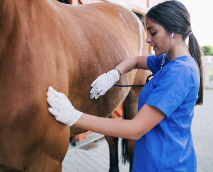
Treatment
It is very important to help maintain circulation during and after the infection.
The swelling that occurs as a result of cellulitis is due to oedema (fluid) build up, which is not only painful in the initial stages, but is often hard to get rid of even once the infection has resolved.
It can lead to continued lameness issues in the affected horse.
Principles of therapy aimed at minimising inflammation, oedema accumulation, and swelling include compression of the leg and cryotherapy.
- Compression (by bandaging the limb) has been shown to be effective in stimulating tissue healing, minimising oedema, and increasing blood flow, providing bandaging is done correctly.
- Cryotherapy (by cold hosing or using ice boots) reduces the inflammatory response in the tissue, reduces the metabolic demand of the tissue, and provides a short-term analgesic effect.
- Walking a horse out in-hand (when they are comfortable enough to) and massage will assist with circulation and encourage fluid drainage.
Vulnerable types
Some horses seem to be more prone to developing cellulitis than others. It is thinner skinned/less hairier breeds that I see most often suffering with the condition.
One of my patients, a retired ex-racehorse called Fin, suffers with several bouts of cellulitis.
His owner is well aware of the typical signs you get with cellulitis and has started physical therapy measures to help alleviate them.
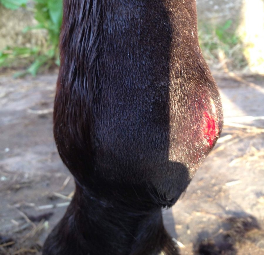
Unfortunately, despite Fin’s owner’s best efforts, on this occasion the heat and swelling doesn’t dissipate sufficiently and serum starts to seep through the horse’s skin.
This combined with an elevated body temperature and the fact that he starts to go off his food means it was time to call a vet and I visit prepared to administer antibiotics and anti-inflammatories.
After careful examination of the leg a small self-inflicted wound is found, which is likely to be the entry point for the infection.
I prescribe Fin five days’ of oral antibiotics and Bute.
Prevention
Two days later, Fin’s owner rings to say the leg has returned to normal and I advise that it is very important that the horse finishes the full course of treatment.
I also advise the following measures to prevent further bouts of cellulitis. These apply to all horses and their owners:
- Maintain a regular exercise programme or plenty of daily turnout as long as the horse is fit to do so, as this helps improve fluid drainage through the lymphatic system from the legs.
- Keep your legs clean and dry, as loss of skin integrity (ie weakening it) is an important risk factor for cellulitis.
- Items used for bathing horses, such as sponges, can carry bacteria that causes bacteria, and so hygienic handling of these things and thorough disinfecting after is important.
- Washing legs can also predispose the skin to chapping, and careful drying of limbs after bathing is essential.
- Avoid using harsh disinfectants like chlorhexidine.
- Don’t turn out in fields/areas that have standing water, mud, or sand. Ideally, find somewhere that doesn’t have this.
- Observe your horse closely and communicate with your vet for early detection of reoccurrence of cellulitis and prompt initiation of treatment.
- Use protective leg boots like brushing boots when you are riding to help lower the risk of wounds and scratches to the lower leg.
Key message
The key message when discussing cellulitis in horses is that time is of the essence.
Do all you can to prevent it occurring, but as soon as you suspect it — or are at a loss to explain what is wrong with your horse’s leg — contact your vet for advice immediately.